CIMM: dental imaging in the Principality of Monaco
Dental imaging especially allows our dental surgeons to analyse and diagnose oral diseases and lesions, or visualise your teeth, gums, jaw and the entire surrounding bone environment.
At our radiology centre, we attach great importance to hygiene and take pride in using top-quality equipment, with high-end machines and the latest technology, with this extending to all our other services, such as bone densitometry, ultrasound or digital radiology.
Do you have any questions? Any doubts? Contact us now for more information, or to schedule an appointment directly at our centre in Monaco.
The different dental imaging techniques performed at the Monaco centre
N.B.:
All these techniques use X-rays, so it is very important to talk about any pregnancy or delayed periods before starting the examination.
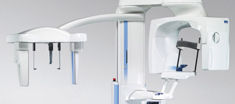
The panoramic dental X-ray
The panoramic dental X-ray is an X-ray providing an overview of all the teeth, as well as the jaws.
This is a very simple and fast examination: the patient just needs to stand in front of the panoramic dental X-ray, and bite down on a small stem, which allows the teeth to be clearly identified. Patients can choose whether to be in a standing or sitting position. The X-ray only takes a few seconds.
Profile or frontal teleradiology
Profile or frontal teleradiology involves an X-ray of the patient’s entire head. This X-ray is often requested during orthodontic treatment.
As with the panoramic dental X-ray, the patient stands or sits in front of the device, and then tips are placed inside the ears and in front of the forehead to secure the head, which must remain stationary. Once again, this is a very fast and simple X-ray.
The Dental Cone Beam
Halfway between the panoramic dental X-ray and the dentascan, the dental Cone Beam is an examination that carries out localised analysis of the teeth.
Patients should be seated with their head wedged in a head support with their mouth closed. Swallowing should be avoided during the examination, which lasts about one minute. We can use this X-ray to accurately reconstruct the tooth or teeth requested by your dentist, with a very low dose of radiation.
The dentascan
The dentascan, as its name suggests, scans the teeth. The patient lies on the examination table with the head placed in the head support.
The examination lasts 2 to 3 minutes, then we perform high definition reconstructions on the requested dental regions. As a complement to the panoramic dental X-ray, it allows you to assess bone stock, before creating a dental implant, or even extracting the wisdom teeth, in order to determine the position of the teeth in relation to the inferior dental nerve canal.

Looking after you for 40 years

Experienced medical and paramedical team

Equipment at the cutting edge of technology